|
Microscopy for time-lapse imaging
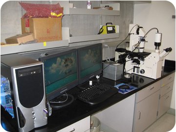
We perform time-lapse imaging in our own lab using a Zeiss axiovert 100 fluroescent microscope (photo below). This microscope is retrofitted with an automated X, Y, Z stages and shutter to generate multiple time-lapse movies using bright light plus green and red fluroescent laser lights. The microscopic observations are captured by a Retiga CCD digital camera, stored on a dedicated Image work station (PC operated by Windows NIT with >100GB disk space) and analyzed using Metamorph and ImageJ image analyses softwares.
We set up multiple in vitro assays and then use the software to mark the position of each ovule (maximum capability = 96) in vitro assays using Metamorph software. Then, we instruct the software to capture a Z stack of images (covering up to 60 microns) of each marked ovule in red, green and bright field channels, once in every 10 minutes for 6-8 hours. The Metamorph software drives all the stages to achieve these tasks and cover 20 ovules in about 10 minutes, then they resume all over again. At the end of the experiment, the time-lapse images for each ovule are assembled using Metamorph and ImageJ softwares. |


